Mapping melanoma
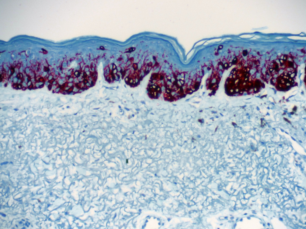
Early detection is crucial for managing any disease. In the case of melanoma, a form of skin cancer that is becoming more prevalent due to climate change and other factors, detection techniques have a long way to go.
The typical method is a biopsy, which can be painful. But alternative approaches that use medical imaging can result in misleading test results, often because they are too narrow in scope.
“There is a serious need for a non-invasive imaging method [that can] scan the entire body for any sign of melanoma,” said Kamran Avanaki, an associate professor in the Richard and Loan Hill Department of Bioengineering.
So, he’s developing one. Avanaki and his team are working on a wide field-of-view optical coherence tomography device that can produce detailed cross-sectional images, allowing for increased specificity and sensitivity—measures for how well positive and negative cases are accurately identified.
“Our hope is that this technique will help lower the number of unnecessary biopsies by pinpointing the most likely malignant lesion on a person with multiple pigmented spots,” Avanaki said. “It will also help lower the cost of diagnosing melanoma by reducing the number of tests the healthcare system needs to perform and lead to earlier detection of the disease.”